 Technology peripherals
 AI
AI
 The Matrix is coming! Burying 10,000 micron electrodes to eavesdrop on the brain, Musk's brain machine will be implanted in the human body
The Matrix is coming! Burying 10,000 micron electrodes to eavesdrop on the brain, Musk's brain machine will be implanted in the human body
There is a complex network in your head - a complex network of 86 billion switches!
Weighs 2 and a half kilograms and consumes only 20W of power, which is equivalent to the energy consumption of a light bulb.
However, it has created infinite miracles in bioelectronics!
The brain is an electronic organ?
The core of brain research is the application of sensors technology.
Whether we are familiar with scalp electrodes, magnetic resonance imaging, or newly pioneered methods such as implanted chips, we are all trying to explore this mysterious organ.
Recently, Imec, a Belgian nanodigital research institute, has pioneered the Neuropixels detector, which is a new probe to observe the living brain at the neuronal level.
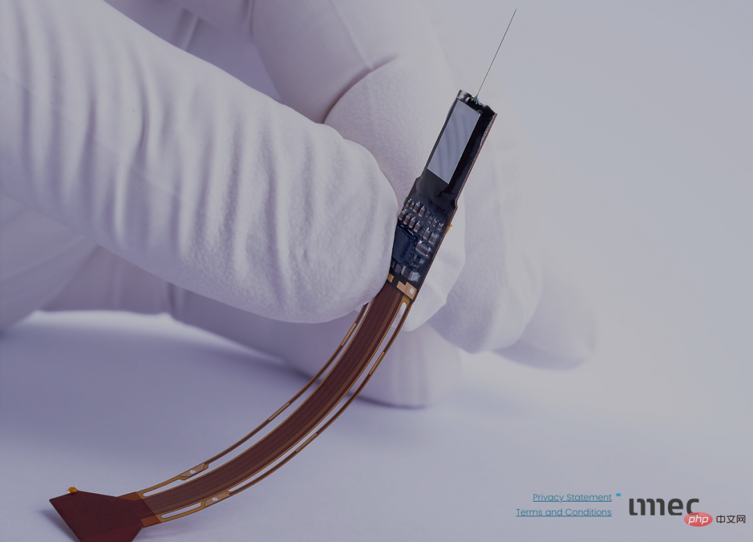
The first-generation Neuropixels detector alone has been delivered to about 650 laboratories around the world. At the same time, Imec also created the OpenScope shared brain observatory to provide open source data to brain researchers around the world.
This is a globally shared neuroscience research facility, equivalent to CERN’s particle accelerator for shared high-energy physics research.
Neuropixel, a new technology for observing brain activity. Its function is similar to imaging, however, it records electric fields instead of light fields.
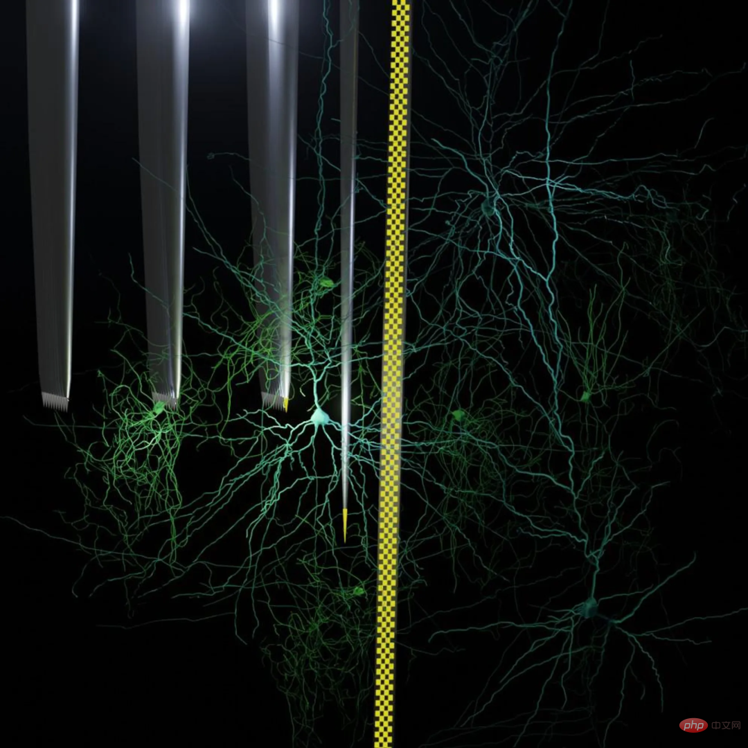
The collaboration began in 2010 with a conversation between engineer Barun Dutta and neuroscientist Timothy D. Harris .
Dutta works at Imec, where he uses state-of-the-art semiconductor manufacturing equipment; Harris works at HHMI (Howard Hughes Medical Institute), where he is a senior neuroscientist.

Dutta brings his knowledge of semiconductors to the field of neuroscience
"We need to study local neural circuits in a freely moving animal, Record the spikes of each neuron," Harris said.

Led by Dutta and Harris, a research team with multidisciplinary backgrounds was formed, including engineers, neuroscientists, software designers and other personnel.
Scientists are exploring how to use advanced microelectronics to invent a new sensor that can simultaneously monitor the electrical conversations between thousands of neurons in any small part of brain tissue.
The system invented by the scientists is named Neuropixels. "Think of us as the Intel of neuroscience," Dutta said. "We provide the chips, and then laboratories around the world use them to write code and do experiments." .
It’s not easy to build a digital probe that is long enough to reach any part of the brain organ, but small enough not to damage delicate tissue on the way in.
In fact, the brain is as elastic as yogurt.
Therefore, scientists need to keep the insertion straight but also allow it to bend inside the shaking brain so that it does not damage neighboring brain cells for a long time.
As the brain guides the body through complex behaviors, detectors need to be durable enough to stay in place and record reliably for weeks or even months.
Neuropixels pushes neuroscience to a higher stage, provides better treatments for brain diseases such as epilepsy and Parkinson's, and paves the way for future brain-computer interfaces.
Back in the 1950s, researchers used a primitive electronic sensor to identify neuronal deactivation in Parkinson's disease patients.
After 70 years of development, with the microelectronics revolution, all components of the brain probe have been miniaturized, and brain electronic sensing technology has made great progress.
In 2021, the system will be upgraded to version 2.0. Compared with the initial version 4 years ago, the number of sensors has been increased by an order of magnitude.
Now, version 3.0 is in the early development stage.
Scientists believe that neuropixels will grow exponentially in accordance with Moore's Law.
And this is just the beginning.
Neuropice2.0!
Biology experts who study the brain suggest that experimenters use gold or platinum for electrodes and then use organometallic polymers for the handles.
However, these materials are not compatible with advanced CMOS manufacturing processes. Therefore, the experimenters conducted some research and did a lot of engineering design. Ultimately, Silke
Musa invented titanium nitride, an extremely strong electrical ceramic that is compatible with CMOS and animal brains.
Also, the material is also porous, which gives it low impedance. Low impedance is very helpful for getting current and clearing the signal without heating nearby cells, creating noise that corrupts the data.
Thanks to extensive materials science research and some related techniques in microelectromechanical systems (MEMS), researchers are now able to control the internal stresses generated during the deposition and etching of silicon rods and titanium nitride electrodes.
In this way, even though the silicon rods are only 23 microns (microns) thick, they can always maintain an almost perfectly straight line.
Each probe consists of four parallel handles, each of which is equipped with 1,280 electrodes. Within 1 centimeter, the probe is long enough to reach anywhere in the mouse brain.
Mouse studies published in 2021 showed that the Neuropice 2.0 device could collect data from the same neurons for six consecutive months while the rodents lived their normal lives.
The elasticity difference between the CMOS-compatible handle and the brain tissue is huge. This leads to a problem: when the probe is in the brain, it inevitably follows the movement of the brain. How should individual neurons be tracked while moving.
We all know that neurons are 20 to 100 microns in size, and each electrode is 15 microns in diameter, which is small enough to record the isolated activity of a single neuron.
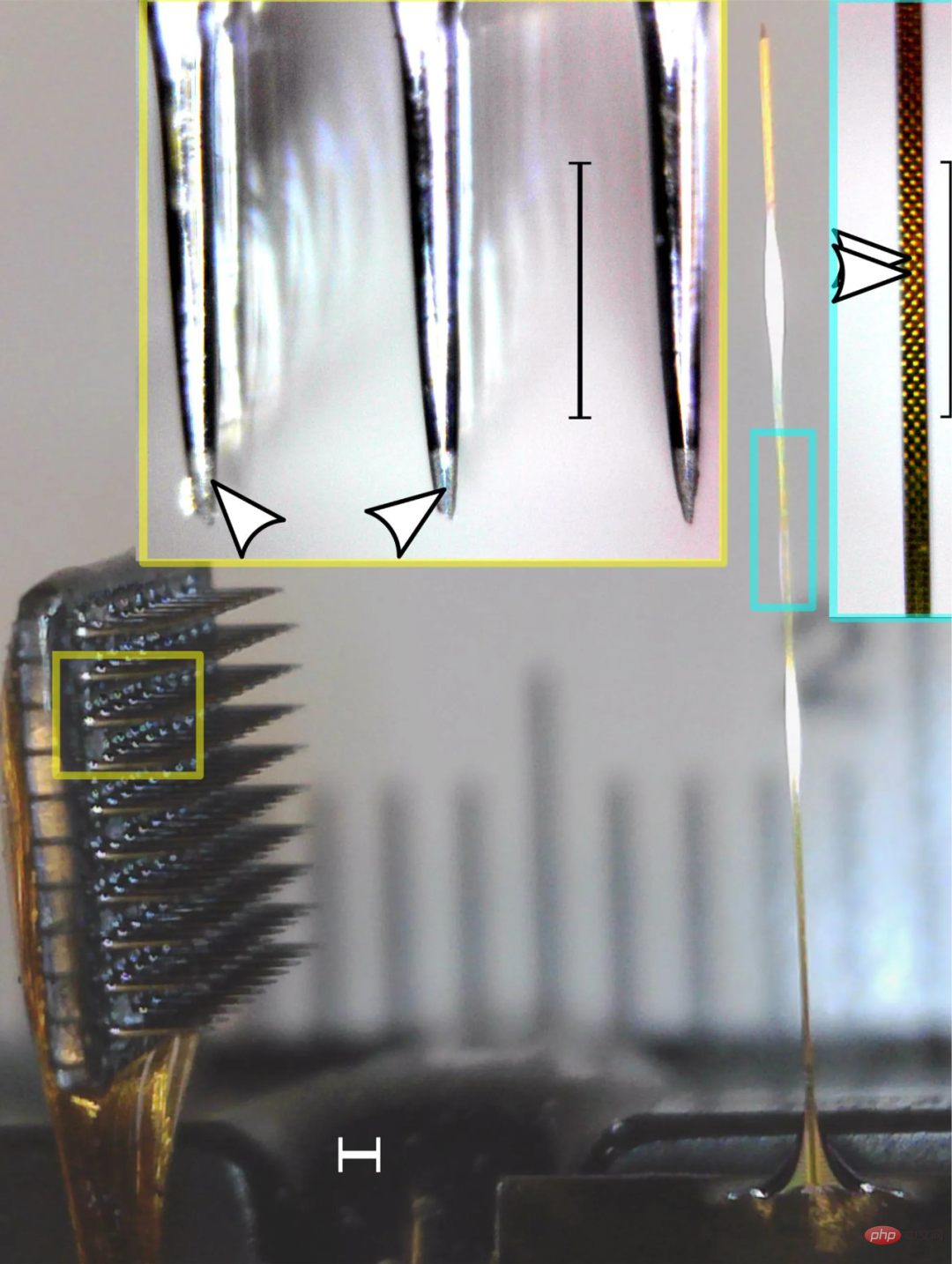
#But after six months of jostling activity, the entire probe may have moved 500 microns within the brain. During this time, any given pixel may see several neurons coming and going.

(Currently the most common neural recording device)
In addition, the 1,280 electrodes on each stem are individually addressable, four The parallel stems give researchers effective 2D readings, which are very similar to images taken by a CMOS camera.
This similarity made the researchers realize that the problem of neuron displacement relative to pixels is very similar to that of the IS system. Like shaking a camera while filming, the neurons in an area of the brain are correlated with their electrical properties.
Researchers can use existing methods to solve the camera shake problem to solve the problem of detection head shaking. With the application of stabilization software, researchers can use automatic correction functions when neural circuits move at will.
Version 2.0 reduces the circuit board located outside the skull, which controls the implanted probe and outputs digital data, to the size of a thumb.
In this way, one circuit board and base can hold two probes, and each probe extends four small handles, with a total of 10,240 recordable electrodes.
The researchers wrote a control program to achieve a high sampling rate and capture a large amount of data. This is 500 times what CMOS imaging chips can usually record. But currently the device can't capture the activity of every neuron it touches.
Continued advances in computer technology will further alleviate existing bandwidth limitations within the next few generations.
In just four years, researchers have almost doubled the density of pixels, doubled the number of pixels that can be recorded simultaneously, and increased the overall number of pixels by more than ten times. The size of external electronic equipment has not increased but decreased, shrinking by half.
The next generation version 3.0 is also under development and will be released around 2025, maintaining a release rhythm of every four years. In version 3.0, the researchers expect that the number of pixels will jump again, allowing monitoring of approximately 50,000 to 100,000 neurons.
Meanwhile, the team plans to continue adding detectors and triple or quadruple the output bandwidth and reduce the base bandwidth by a factor of two.
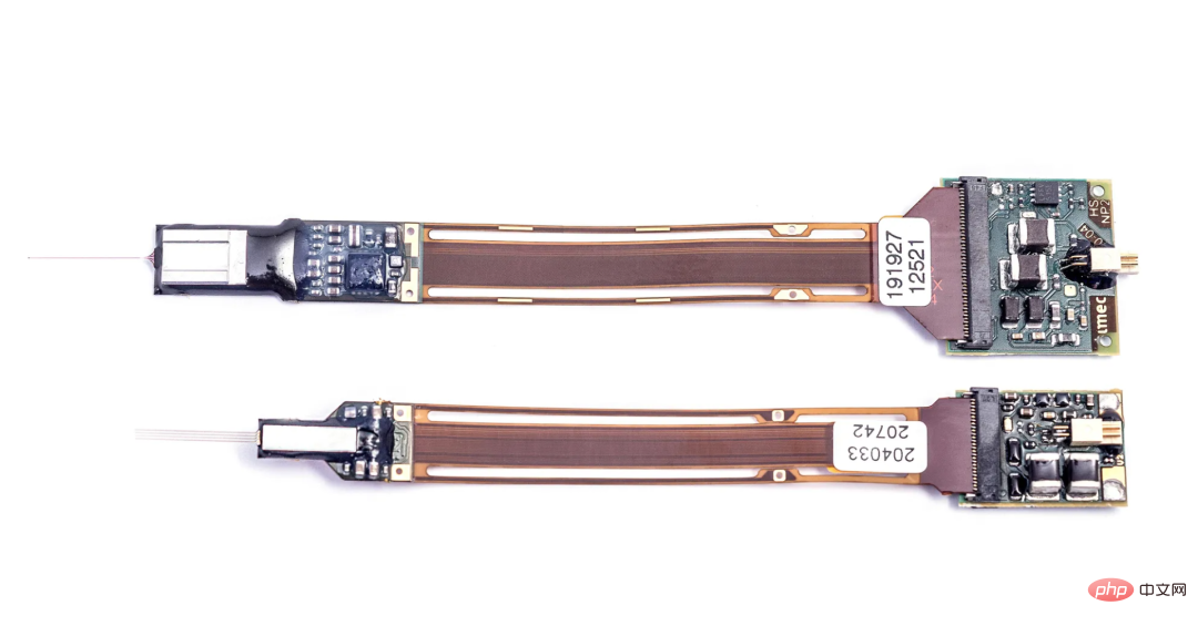
(The first Neuropion device. There are 966 electrodes on the handle.)
Frankenstein opened the skull, the first human brain machine
In order to advance scientific research, many Frankensteins have conducted experiments on their own bodies. In 2014, Phil Kennedy, a neuroscientist in his late 70s in the United States, sawed open his own skull and implanted electrodes into his brain.
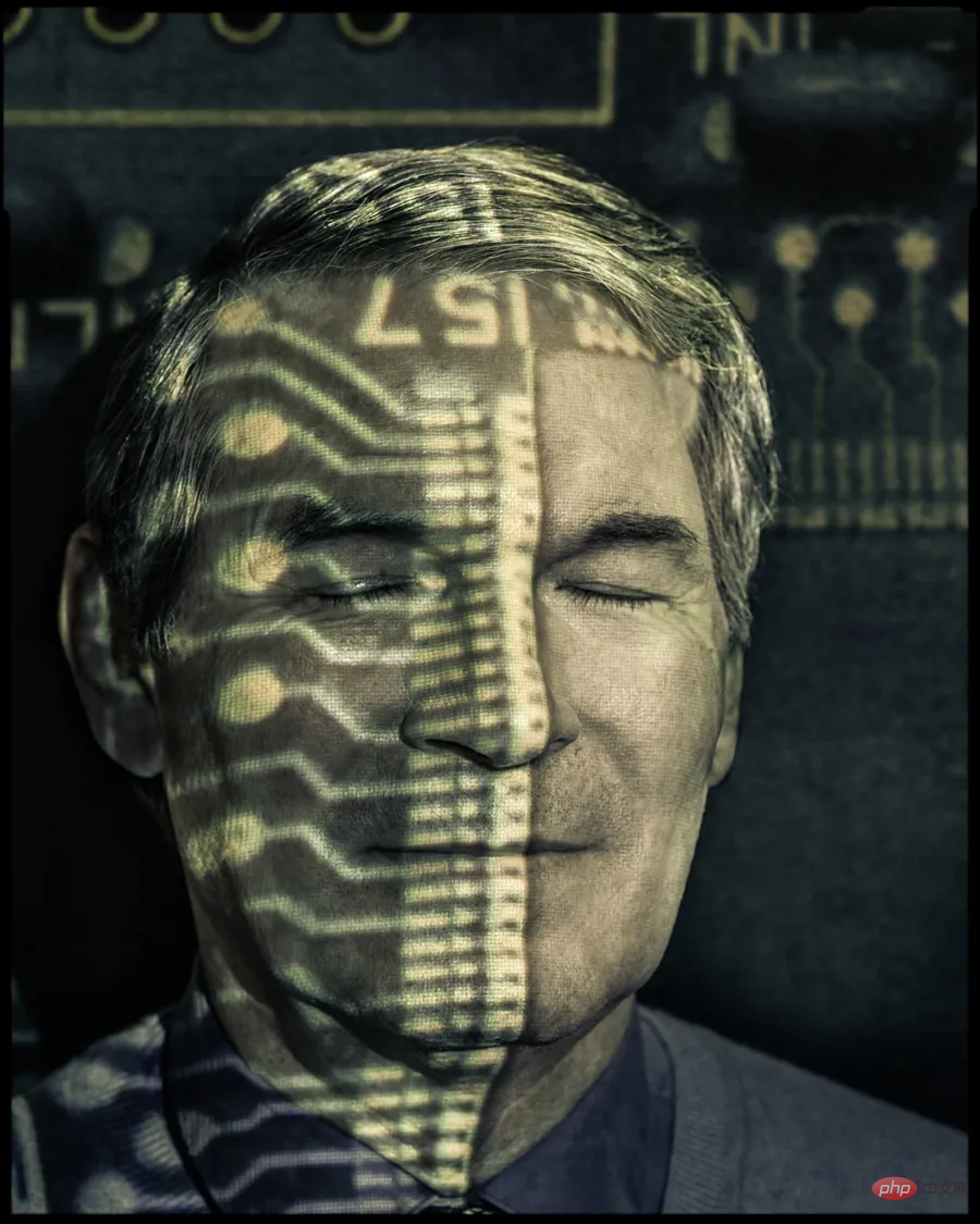 At that time, because he could not find experimental subjects and research funds were about to dry up, Kennedy decided to operate on his own brain. The brain surgery lasted for 11 and a half hours, and it actually didn't go very smoothly.
Kennedy lost the ability to speak when he woke up. He did this to create a speech decoder that would allow patients who cannot speak to "speak" again through a brain-computer interface.
Previously, Phil Kennedy had been researching in this field for nearly 30 years. He was a well-known neuroscientist and was called the "father of cyborgs" by many.
The invasive brain-computer interface he developed in the 1990s allowed a severely paralyzed person to learn to use his brain to control the computer cursor to type, so that others could "hear" his voice.
There are countless studies on brain-computer interfaces, and the most exciting and exciting one is Neuarlink’s research. Just in August 2020, Musk announced Neuarlink's major breakthrough at the press conference.
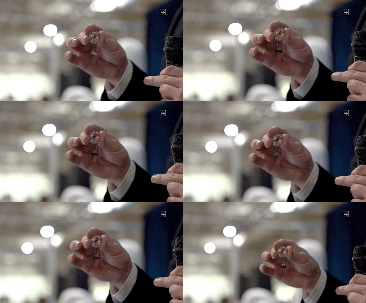
This time, the magical device created by Musk is only the size of a coin. It is surgically implanted in the skull and can be used for a whole day when fully charged. Musk said that the most essential issue of brain-computer interface is "wiring".
The above is the detailed content of The Matrix is coming! Burying 10,000 micron electrodes to eavesdrop on the brain, Musk's brain machine will be implanted in the human body. For more information, please follow other related articles on the PHP Chinese website!

Hot AI Tools

Undresser.AI Undress
AI-powered app for creating realistic nude photos

AI Clothes Remover
Online AI tool for removing clothes from photos.

Undress AI Tool
Undress images for free

Clothoff.io
AI clothes remover

Video Face Swap
Swap faces in any video effortlessly with our completely free AI face swap tool!

Hot Article

Hot Tools

Notepad++7.3.1
Easy-to-use and free code editor

SublimeText3 Chinese version
Chinese version, very easy to use

Zend Studio 13.0.1
Powerful PHP integrated development environment

Dreamweaver CS6
Visual web development tools

SublimeText3 Mac version
God-level code editing software (SublimeText3)

Hot Topics
 1393
1393
 52
52
 37
37
 111
111
 Musk opens a new chapter for Tesla: plans to launch new, lower-cost electric models in 2025
Jan 25, 2024 pm 05:51 PM
According to news on January 25, Tesla CEO Elon Musk revealed during today’s 2023 fourth quarter and full-year results conference call that Tesla is about to usher in an important milestone. Faced with the challenge that the cost of current models is close to the limit, Musk made it clear that Tesla is actively preparing to put into production new models next year. This move is aimed at further improving Tesla's production efficiency and reducing costs to meet growing market demand. Musk also said that Tesla will continue to be committed to innovation and technological advancement to provide consumers with better electric vehicle products. Tesla’s future development prospects are exciting. It is understood that Musk plans to start the production of next-generation electric vehicles at Tesla’s Gigafactory in Texas in the second half of 2025.
 Canada plans to ban hacking tool Flipper Zero as car theft problem surges
Jul 17, 2024 am 03:06 AM
This website reported on February 12 that the Canadian government plans to ban the sale of hacking tool FlipperZero and similar devices because they are labeled as tools that thieves can use to steal cars. FlipperZero is a portable programmable test tool that helps test and debug various hardware and digital devices through multiple protocols, including RFID, radio, NFC, infrared and Bluetooth, and has won the favor of many geeks and hackers. Since the release of the product, users have demonstrated FlipperZero's capabilities on social media, including using replay attacks to unlock cars, open garage doors, activate doorbells and clone various digital keys. ▲FlipperZero copies the McLaren keychain and unlocks the car Canadian Industry Minister Franço
 Microsoft, OpenAI plan to invest $100 million in humanoid robots! Netizens are calling Musk
Feb 01, 2024 am 11:18 AM
Microsoft and OpenAI were revealed to be investing large sums of money into a humanoid robot startup at the beginning of the year. Among them, Microsoft plans to invest US$95 million, and OpenAI will invest US$5 million. According to Bloomberg, the company is expected to raise a total of US$500 million in this round, and its pre-money valuation may reach US$1.9 billion. What attracts them? Let’s take a look at this company’s robotics achievements first. This robot is all silver and black, and its appearance resembles the image of a robot in a Hollywood science fiction blockbuster: Now, he is putting a coffee capsule into the coffee machine: If it is not placed correctly, it will adjust itself without any human remote control: However, After a while, a cup of coffee can be taken away and enjoyed: Do you have any family members who have recognized it? Yes, this robot was created some time ago.
 Hacker releases jailbreak tool compatible with iOS 15 and iOS 16
May 29, 2023 pm 04:34 PM
Apple has been working hard to improve the security of its operating system and devices, which has been proven considering how difficult it is for hackers to create jailbreak tools for iOS 15. But those who are keen on tinkering with iOS can now celebrate, as the palera1n team has released a jailbreak tool that is not only compatible with iOS15, but also iOS16. For those unfamiliar, the jailbreaking process removes software restrictions on an iOS device so that users can access and modify system files, allowing for various modifications such as tweaks, themes, and sideloading of apps outside of the App Store. Of course, Apple has always opposed the process of jailbreaking its devices. iOS15 and iOS16 jailbreak paler
 Musk satirizes artificial intelligence AI hype: The essence of 'machine learning' is statistics
Jun 13, 2023 pm 12:13 PM
Drive China News on June 12, 2023 Recently, Tesla CEO Elon Musk posted a picture on Twitter on Saturday, which seemed to be a satire on the current hype about "artificial intelligence". The picture shows a passerby wearing a "Machine Learning" mask. When he took off his mask, a face with "Statistics" written on it appeared. The essence of artificial intelligence, which means the current fire, is the result of data statistics. It is worth noting that there are probably not a few technology leaders who hold such opinions. Musk had previously signed an open letter with Apple co-founder Steve Wozniak and thousands of AI researchers, calling for a suspension of research on more advanced AI. AI technology. However, this letter was questioned by many experts and even the signers
 Tesla will support Apple Watch to control cars, Musk personally confirms
Mar 07, 2024 pm 01:19 PM
Recently, Tesla CEO Musk responded positively to whether Tesla will increase integration functions with Apple Watch. He said that Tesla owners can expect to use Apple Watch to control their cars, which received widespread attention. However, the specific implementation timetable for this feature has not yet been announced, making people's expectations for the future even higher. It is understood that Tesla’s Apple Watch app plans to provide a series of practical functions, including unlocking the car, preheating or precooling the interior environment, enabling or disabling Sentry mode, and remotely locking or unlocking the car. These features will greatly enhance the convenience and experience of Tesla owners. Currently, there are some third-party applications in the AppStore, such as Tess
 SpaceX Starship Base adds new tower, one step closer to Musk's dream of Mars
Aug 23, 2024 am 07:33 AM
According to news from this site on August 22, SpaceX officially announced that the second starship launch tower at its Starbase (i.e. South Texas launch site) has now been capped (completed all stacking of the tower). This process only requires 41 days. fenyeSpaceX also said that they are preparing to launch two manned Dragon spacecrafts within the next month to carry out SpaceX's 14th and 15th manned missions, namely the "Polaris Dawn" commercial space mission and NASA's Crew- 9 tasks. Among them, the "Polaris Dawn" mission will complete mankind's first commercial spacewalk and break the record for the highest orbital altitude of a manned mission in low-Earth orbit. Details can be found in previous reports on this site. Furthermore, if Boeing cannot pass its own
 The number one Blizzard player in history and the richest man in the world also collapsed and bluntly said he would give up Blizzard games.
Feb 07, 2024 am 09:12 AM
The Light of Technology, Iron Man, and the World's Richest Man are all one of the many shining labels on Musk. In fact, Musk also has another label, which is that he is an avid fan of Blizzard games. Even though he runs some of the most important companies in the world, Technology company, Musk will still spend a lot of time playing Blizzard games. After Blizzard launched the new game Diablo 4 in 2023, Musk was full of praise for it, even though this game was abandoned by most old players. But the recent poor performance of Diablo 4 Season 3 has made Musk unable to hold back. How obsessed is Musk with playing Blizzard games? Xiaotan will give you a few examples first. On October 2 last year, Musk’s X (twitter) platform launched the live broadcast function, and Musk led the live broadcast debut, using explosives to play Diablo 4.


